
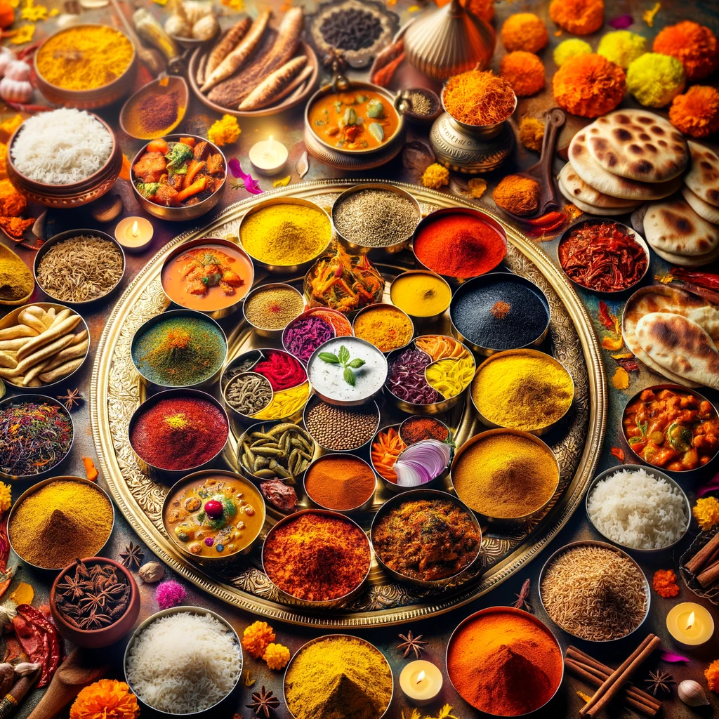
Week Four: Indian Cuisine - Spicing Up Health and Vitality 🍛🌶️
Welcome to Week 4 of Mindful Diabetes Inc.'s inspired series, "Plant-Based Strategies for Health and Wellness." As we deepen our culinary voyage, we embrace the rich tapestry of Indian cuisine, renowned for its intricate spices and vibrant flavors that not only please the palate but also promote diabetes management and cognitive health.
Week Four: The Healing Spices of India 🌿💚
This week, we explore the aromatic world of Indian cooking, highlighting ingredients that have been used for centuries not only for their taste but also for their anti-inflammatory properties. Indian cuisine offers a cornucopia of plant-based delights that can enhance gut health and support a balanced Diabetes Microbiome. Learn how traditional dishes, adapted to a healthful eating plan, can become a staple in your mindful diet.
Incorporating Tradition into Modern Wellness Practices 🥘✨
We will guide you through the essentials of crafting an Indian-inspired pantry, rich in legumes, whole grains, and spices like turmeric, cumin, and coriander. These ingredients are key in creating meals that are not only authentic but also align with our mission to support wellness through diet.
Embark on this week's delectable journey, where the spices of India meet the science of health. Our series continues to illuminate how diverse cuisines can enrich our approach to diabetes care and Alzheimer's prevention.
Stay tuned as we unlock the secrets to blending traditional Indian flavors with the principles of mindful eating. Together, let's savor each dish and celebrate the harmony of taste and health that Indian cuisine brings to our tables.

Support prevention education
Your gift supports Mindful Diabetes education, tools, and prevention-focused community work.
Your Indian Cuisine Immersion: Week 4 of Mindful Eating 🍛🌿
Welcome to the vibrant Week 4 of our "Plant-Based Strategies for Health and Wellness" series at Mindful Diabetes Inc., where we explore the aromatic and healthful world of Indian cuisine. This week we dive into the spices and flavors of India, renowned for their diabetes-friendly benefits and brain-boosting properties.
Embracing Indian Cuisine for Diabetes 🇮🇳💡
Indian cuisine, with its rich tapestry of herbs and spices, is perfectly poised to support a diabetes-conscious diet. Turmeric, cumin, fenugreek—these are more than just flavor enhancers; they are part of a powerful arsenal in blood sugar management and cognitive health maintenance.
Creating a Diabetes-Friendly Indian Pantry 🛍️🥘
Building an Indian-inspired pantry is a delicious adventure in itself:
Whole Grains: Choose fiber-rich options like brown basmati rice and whole wheat chapatis to aid in slow sugar release and satiety.
Legumes Galore: Lentils and chickpeas are not only staple proteins in Indian Food For Diabetes but also great sources of fiber and micronutrients.
Spice it Up: Incorporate a variety of spices such as turmeric, which is linked to anti-inflammatory properties beneficial for managing Diabetes and Indian Cuisine.
Healthy Fats: Don't forget to include heart-healthy fats found in nuts, seeds, and oils, which are essential for hormone health and cell function.
This week, let Indian Cuisine for Diabetes guide your meal planning. We'll be sharing recipes that bring traditional Indian dishes into your diabetes management plan, infusing each bite with both culture and care.
Stay tuned for flavorful dishes that embrace the essence of Indian cuisine while keeping your health in check. From the tandoor to the table, let's celebrate the joy of Indian cuisine and its place in our mindful eating journey. 🌶️🍲
Free wellness tools
Choose a tool for your next healthy step
Explore AI-guided learning, daily habit tracking, and a playful nutrition game built around Mindful Diabetes prevention education.
Daily habits app
Want one simple place to practice the habits in this article? Memovela is available on the App Store and on the web.

🌿 Produce:
- 🍊 2 oranges
- 🍎 1 apple
- 🍍 1 pineapple
- 🥭 1 unripe mango
- 🍇 1 grapefruit
- 🍓 1 large container fresh strawberries
- 🌿 1 cup cilantro
- 🍃 1 cup mint leaves
- 🌶️ 3 jalapeno
- 🥦 1.5 lbs fresh spinach (plus more for lunch salad)
- 🧅 1 red onion
- 🍋 2 lemon
- 🧄 1 garlic bulb
- 🥬 1 large sweet potato
🥖 Grains and Beans:
- 🌾 1 cup black lentils*
- 🥘 12 oz firm tofu
- 🥣 15 oz can chickpeas*
- 🍚 1 lb brown rice*
- 🍞 3 whole wheat naan
- 🥣 1.5 c old fashioned oats*
🥛 Dairy and Non-Dairy:
- 🥛 1 1/2 cup plain yogurt
- 🥛 5 oz low fat milk or unsweetened almond milk
- 🫐 1/2 c frozen wild blueberries*
🥄 Pantry and Misc.:
- 🍅 4 oz can diced tomatoes*
- 🥭 1 oz mango puree
- 🍫 1 cup shelled pistachios
- 🍫 5 oz dark chocolate
- 🥥 4 oz dry shredded coconut
- 🥜 1/4 c walnuts*
- 🥜 1/2 c almonds*
- 🌰 2 T chia seeds
- 🍂 1-2 T seasoning (optional; e.g., taco seasoning, Old Bay, chili powder)
- 🧂 1.2 T cumin*
- 🌿 1/4 t cardamom
- 💛 1/2 t turmeric
- 🌾 1/2 T coriander
- 🌶️ 1/2 t chili powder*
- 🌶️ 2 whole dried red chili peppers
- 🌿 1/4 t cornstarch
- 🌿 1/2 t fennel seeds
- 🌶️ 1/4 t cayenne*
- 🍯 1 T granulated sugar*
- 🌶️ 1-2 t chaat masala
- 🌿 1 bay leaf
- 🛢️ 2 T canola oil*
- 🫒 Olive oil*
- 🍾 3 T vinegar*
- 🍯 1 t honey*
By adding these items to your pantry, you're well on your way to crafting delicious Indian-inspired meals that are not only delightful but also support diabetes management and Alzheimer's prevention. Remember, every spice, every grain, and every piece of produce is not just food—it's fuel for wellness and a dash of delight for your senses. Happy cooking! 🍳✨
*Items marked with an asterisk indicate ingredients that may already be in your pantry. Make sure to check for items that need to be replenished before heading to the grocery store. Happy cooking! 🛒👩🍳

Your Two Day Meal Plan for a Healthy Diet
🌅 Day 1: A Mindful Start with Indian Flavors
Breakfast: Begin with warmth and spice:
- 🥣 Oatmeal stirred with turmeric and a dash of cinnamon for anti-inflammatory benefits. Sprinkle with antioxidant-rich walnuts and a swirl of honey for sweetness.
Lunch: Midday vibrancy with a touch of tradition:
- 🥙 A lentil salad with whole wheat naan. Add a dollop of green chutney for a tangy twist.
Dinner: Savor the richness of Indian spices:
- 🍲 Chana masala paired with roasted cauliflower for a fulfilling, fiber-rich evening meal.
Snack: Refresh and hydrate:
- 🍓 Enjoy a simple fruit salad with a sprinkle of chaat masala for an unexpected kick, alongside calming turmeric tea.
🌄 Day 2: Continuation of Flavorful Nourishment
Breakfast: A sweet and nutritious start:
- 🍓 Overnight oats with a topping of sliced strawberries a la lemington for a fruity beginning.
Lunch: A light yet satisfying midday meal:
- 🌯 Chana masala and roasted cauliflower served on fresh spinach leaves, scooped with a piece of whole wheat naan for added texture and fiber.
Dinner: An evening indulgence in creamy comfort:
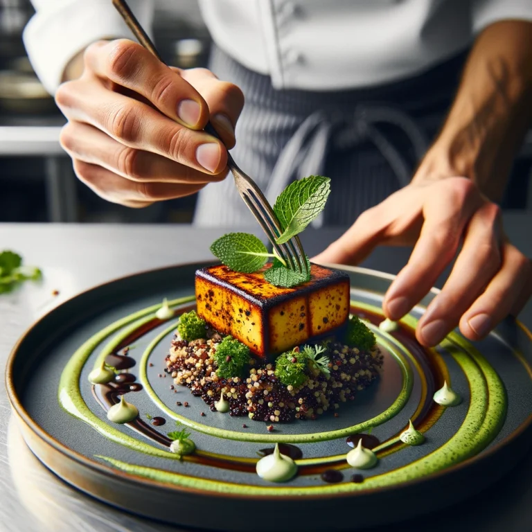
- 🥘 Saag paneer with tofu, a creamy spinach dish that's both comforting and packed with protein, served with whole wheat naan.
Snack: A zesty and refreshing end to your day:
- 🍊 Orange slices dusted with chaat masala and a sprinkle of lime for an energizing and flavorful snack.
Enjoy these meals, which are infused with the essence of Indian cuisine, offering you a balance of spices, herbs, and whole foods for a diabetes-conscious diet. Take pleasure in each aromatic bite and let these meals nourish your body and delight your palate. 🌿🥘✨

Savoring the Spice of Life: Closing Week 4 with Indian Cuisine 🍛🌿
As we conclude this vibrant chapter of our gastronomic journey, Week 4 has immersed us in the aromatic and wholesome world of Indian cuisine. At Mindful Diabetes Inc., we've embraced the rich tapestry of spices and flavors that characterize Indian cooking, showcasing how it can support diabetes management and cognitive health.
Infusing Wellness with Indian Spices 🌶️
Our "Pathways to Wellness" series has explored how the traditional foods of India can enhance a diabetes-conscious diet. We've learned that the complex spices and diverse ingredients of Indian recipes aren't just for pleasing the palate; they're also allies in maintaining blood sugar levels and supporting brain health.
Culinary Exploration Meets Healthful Innovation 🥘🥦
This week has been a testament to the adaptability of Indian cuisine to health-conscious lifestyles. By focusing on fiber-rich legumes, antioxidant-packed spices, and nutrient-dense vegetables, we've seen how Indian dishes can be both nourishing and indulgent.
Looking ahead, Mindful Diabetes Inc. remains dedicated to uncovering more culinary secrets that benefit both body and mind. As we anticipate the next installment of our series, we remain committed to guiding you through the delicious possibilities of mindful eating.
Let's carry forward the essence of Indian cuisine, where every spice and ingredient plays a role in crafting a balanced and healthful diet. Together, let's continue this flavorful quest, weaving the joy of Indian cooking into our daily lives for better health and vibrant wellness. 🌟🍲
📖 Continue Exploring 🌐
Don’t stop here! Dive deeper into the fascinating connection between mental wellness and physical health in our upcoming blogs. There’s always more to discover and explore.
💌 Stay Informed and Inspired 📬
Subscribe to our newsletter for the latest insights, tips, and updates on the mindful path to better health. Together, we’ll continue to unlock the secrets of a balanced and vibrant life.
Thank you for your continued support and readership. Here’s to a healthier, happier you! 🌈💚
Interested in staying up to date on the latest from Mindful Diabetes and our Type 2 & Type 3 Diabetes Guide? Subscribe to our newsletter!
Facebook Instagram Youtube LinkedinCheck out our Shop Over on Tik Tok!⬇️