

Why Amyloid Still Matters, and Why It Is Not the Whole Disease
Few words in Alzheimer’s research carry as much history as amyloid. Amyloid beta is a small protein fragment that can collect outside neurons and form the plaques that appear in many Alzheimer’s disease brains. Those plaques are important. They are visible, measurable, and biologically meaningful. But they are not the entire disease, and they are not a simple scoreboard for memory.
As of January 15, 2026, the most useful amyloid research is becoming more precise and more honest at the same time. Researchers are no longer asking only whether plaques are present. They are asking where plaques sit, what they are made of, how old they are, how nearby cells respond, what tau is doing, whether blood vessels are stressed, whether inflammation is helping or harming, and whether a biological improvement in the brain changes the life of a person sitting across from a clinician.
This article belongs beside our Mindful Diabetes conversations about Type 3 Diabetes, the diabetes-Alzheimer’s connection, insulin resistance and cognitive decline, and glucose metabolism and brain health. The goal is not to turn amyloid into a villain or a hero. The goal is to understand what this biology can tell us, what it cannot tell us, and why the next generation of Alzheimer’s research is becoming more nuanced.
For helpful background, the National Institute on Aging explains how Alzheimer’s disease involves amyloid plaques, tau tangles, inflammation, vascular changes, and progressive brain-cell injury, while its overview of Alzheimer’s causes and risk factors places amyloid within a larger mix of age, genes, vascular health, metabolism, and environment.
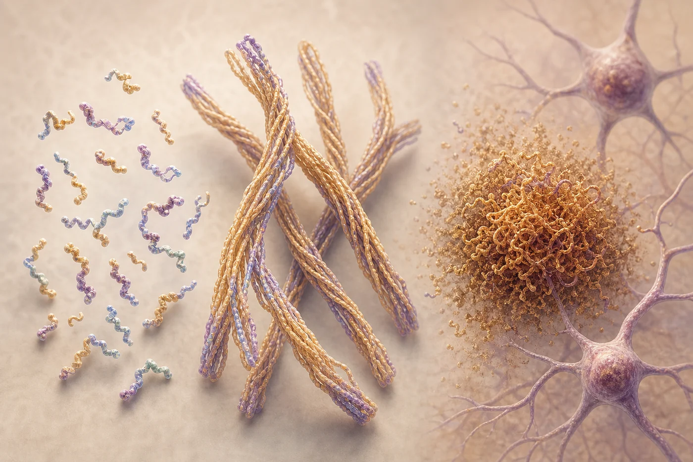
How Plaques Form Is More Complicated Than a Pileup
Amyloid plaques are often described as deposits, but that word can make them sound like dust on a shelf. The biology is more active than that. Amyloid beta fragments can exist as smaller soluble assemblies, longer fibril-like structures, and larger plaques embedded in a living tissue environment. Each form may interact differently with neurons, glia, synapses, blood vessels, and immune cells.
One helpful way to understand the field is to separate what can be seen from what may be doing damage. Plaques are visible with staining and imaging tools. Soluble amyloid assemblies are harder to track, yet many researchers suspect they may stress synapses and cell signaling in ways that a plaque count alone cannot capture.
That is one reason scientists care about plaque structure and composition. A 2025 Nature Communications study used chemical imaging to show that amyloid plaques are not all chemically identical across the Alzheimer’s disease spectrum. The paper, Chemical imaging delineates Aβ plaque polymorphism across the Alzheimer’s disease spectrum, helps explain why two people can both have amyloid pathology while still showing different patterns of disease biology.
Another 2025 Nature Communications study followed plaque maturation in an Alzheimer’s mouse model with isotope-encoded spatial biology. The study, Isotope-encoded spatial biology identifies plaque-age-dependent maturation and synaptic loss in an Alzheimer’s disease mouse model, points toward a sharper question: not simply “How much plaque is there?” but “What kind of plaque is present, how mature is it, and what is happening to nearby synapses?”
Location May Matter as Much as Quantity
The brain is not a uniform container. A plaque near a vulnerable memory network may not mean the same thing as a plaque in a less clinically sensitive region. A compact plaque surrounded by activated microglia may have a different biological neighborhood than a diffuse deposit sitting near stressed blood vessels or fragile synapses.
In practice, this is why modern Alzheimer’s research is moving away from single-number thinking. A person’s amyloid PET scan, tau pattern, cognitive testing, vascular history, glucose control, sleep, medications, and symptoms all contribute context. The plaque signal matters, but its meaning changes depending on the rest of the map.
This also helps explain why our older posts on blood sugar and the body, daily wellness habits, and walking for health remain relevant. They do not promise to remove plaques. They help people think about the whole body environment that supports the brain: circulation, inflammation, stress, sleep, glucose stability, and cardiovascular resilience.
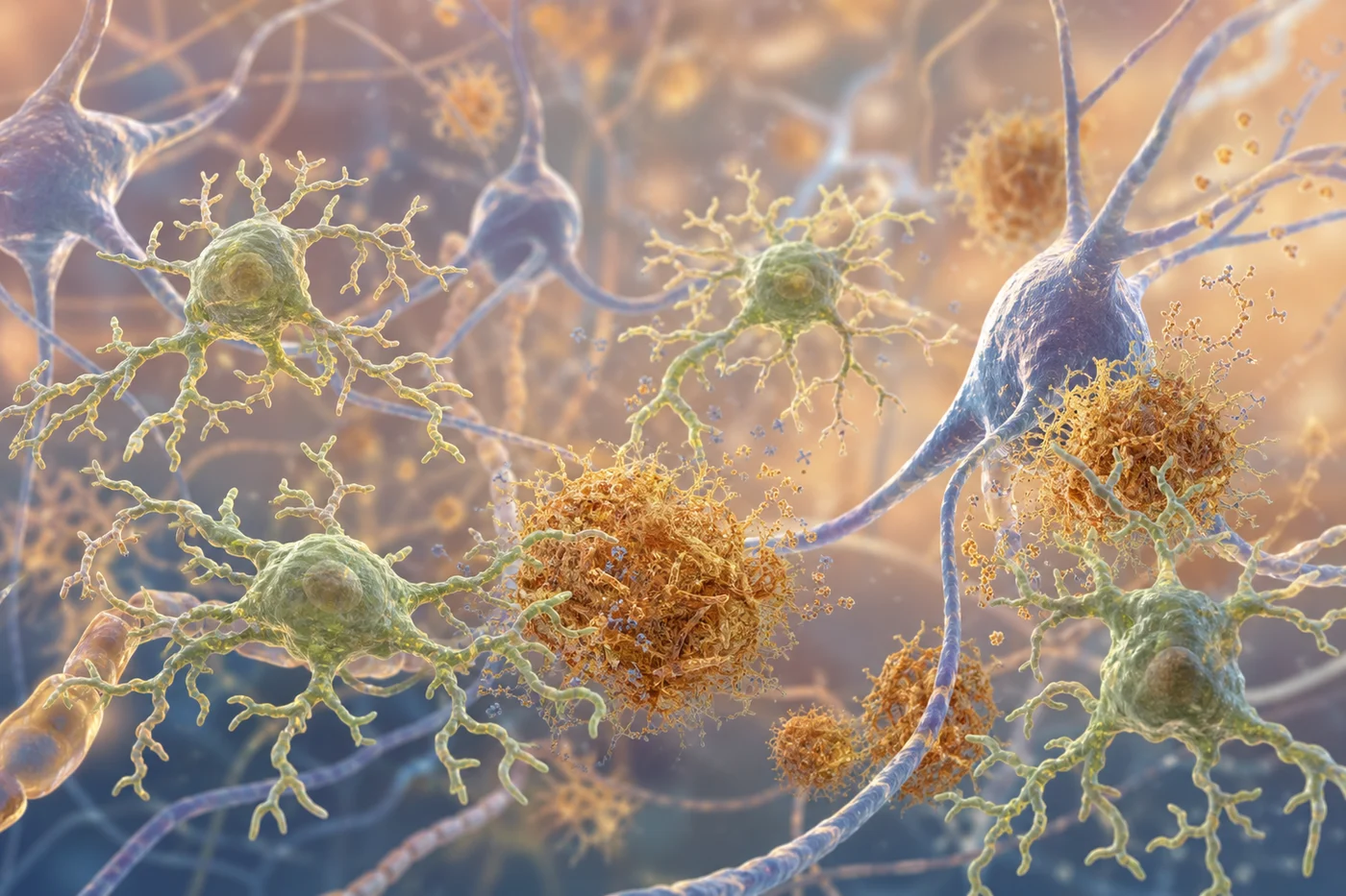
The Immune System Is Not Just Cleaning Up Debris
Microglia are the brain’s resident immune cells. They can survey tissue, respond to injury, process cellular debris, and interact with amyloid deposits. Around plaques, microglia may help compact amyloid, limit spread, or clear material. They may also contribute to inflammation, synaptic stress, and local tissue changes depending on timing, genetic background, and disease stage.
A 2025 Nature Medicine paper studied human Alzheimer’s disease brains from immunized patients and described microglial mechanisms involved in amyloid beta clearance. The study, Microglial mechanisms drive amyloid-β clearance in immunized patients with Alzheimer’s disease, is important because it looks beyond the idea that antibodies simply “remove plaque.” It asks which cells participate, what tissue response follows, and why immune biology may determine whether amyloid clearance is helpful, incomplete, or potentially risky.
The practical significance is that the brain’s immune response is not an accessory detail. It may shape whether amyloid-targeting strategies are safe, how plaques change, and how nearby neurons respond. This is one reason researchers continue studying APOE, complement signaling, microglial states, vascular amyloid, and inflammation as part of the amyloid story rather than as separate side topics.

Seeing Plaques More Precisely Does Not Mean Seeing Everything
Amyloid PET imaging changed Alzheimer’s research because it allowed scientists to estimate amyloid plaque burden in living people rather than only after death. That made clinical trials more targeted: researchers could enroll people with evidence of amyloid biology and measure whether a treatment changed that signal over time.
But amyloid PET has boundaries. It mainly reflects fibrillar amyloid plaque burden. It does not directly measure every soluble amyloid species, every form of tau, every inflammatory state, or every cause of cognitive change. A scan can clarify biology, but it cannot replace the person’s story, cognitive testing, medical history, neurological exam, MRI context, or caregiver observations.
The updated Appropriate Use Criteria for Amyloid and Tau PET, published in January 2025 by an Alzheimer’s Association and Society of Nuclear Medicine and Molecular Imaging workgroup, reflects this more careful era. Amyloid and tau PET can be powerful when the clinical question is clear. They are less helpful when used without a thoughtful reason for testing or a plan for explaining the result.
That same caution applies to blood biomarkers. Blood-based tests are becoming more useful in research and specialty settings, especially when paired with other data. Still, a biomarker is evidence, not destiny. It should make the conversation more precise, not more frightening.
What Anti-Amyloid Treatments Have Taught the Field
Anti-amyloid antibodies have changed the Alzheimer’s field because they showed that plaque biology can be modified in people. Lecanemab and donanemab both reduced amyloid and slowed clinical decline in selected groups with early Alzheimer’s disease, confirmed amyloid pathology, and careful monitoring. The lecanemab trial is published as Lecanemab in Early Alzheimer’s Disease, and the donanemab trial is published as Donanemab in Early Symptomatic Alzheimer Disease: The TRAILBLAZER-ALZ 2 Randomized Clinical Trial.
At the same time, these medicines have taught researchers to be humble. Slowing decline is not the same as restoring lost memory. Reducing plaques is not the same as reversing Alzheimer’s disease. Treatment benefit depends on stage, biology, risk, access, monitoring, and the outcome being measured.
Other trials underline that point. In two phase 3 trials of gantenerumab in early Alzheimer’s disease, amyloid was lowered but the clinical endpoints were not met. In the A4 Study, solanezumab in preclinical Alzheimer’s disease did not slow cognitive decline. These results do not make amyloid irrelevant. They show that the right target, the right timing, the right population, and the right biological mechanism all matter.
Safety also belongs in the center of the discussion. The FDA prescribing information for Leqembi and Kisunla includes warnings about amyloid-related imaging abnormalities, often called ARIA. ARIA can involve brain swelling or bleeding changes seen on MRI. Many cases are asymptomatic, but some can be serious. That is why treatment decisions require medical supervision, MRI monitoring, risk assessment, and a clinician who can explain the tradeoffs.
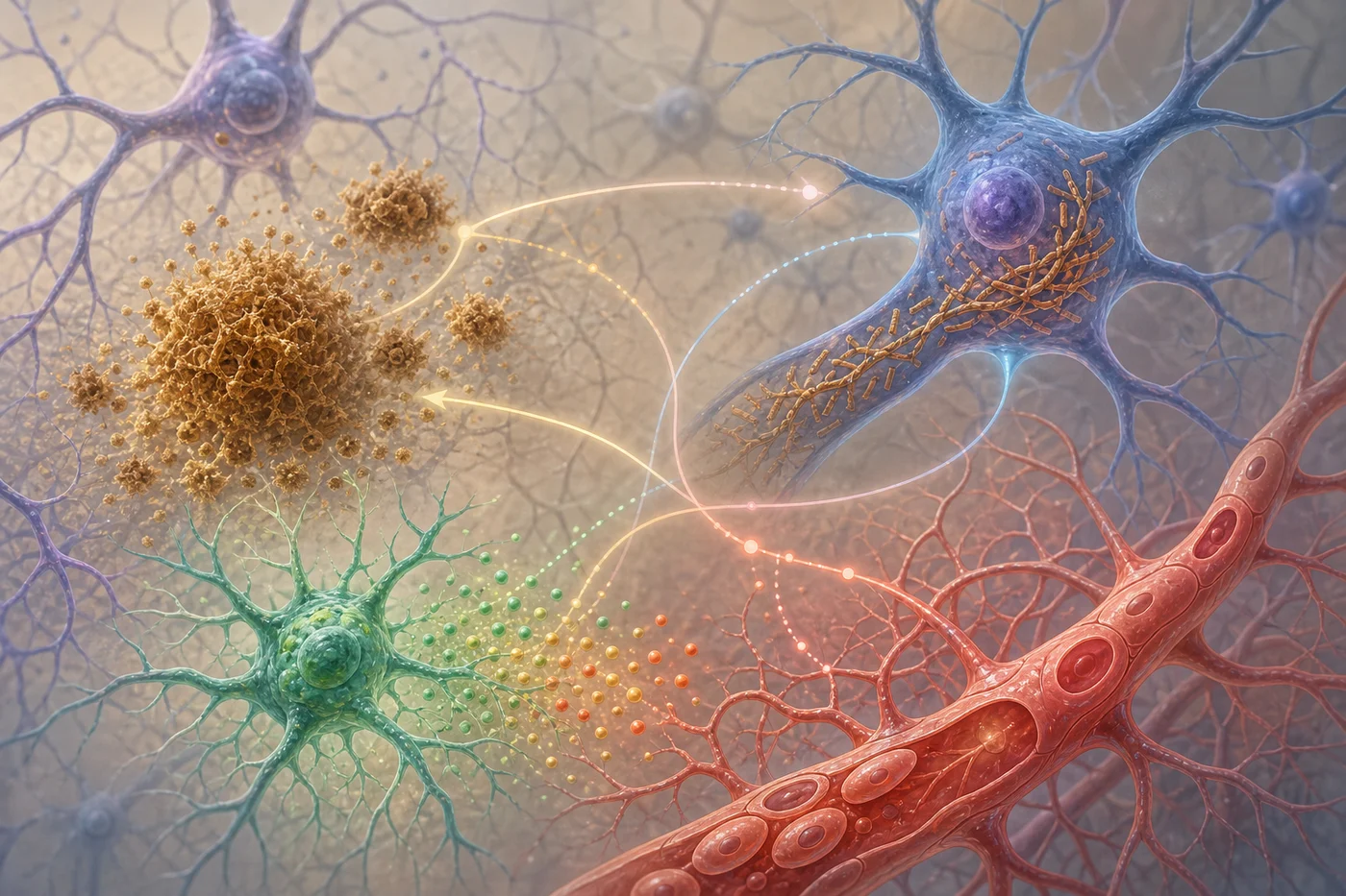
Amyloid, Tau, Inflammation, and Blood Vessels Travel Together
Amyloid plaques may arrive early in the Alzheimer’s timeline, but tau pathology often lines up more closely with symptom patterns and neurodegeneration. Inflammation can be protective in one context and damaging in another. Blood vessels influence oxygen delivery, waste clearance, blood-brain barrier function, and the brain’s ability to recover from stress. Metabolic health shapes the environment in which all of this biology unfolds.
This is why the Mindful Diabetes lens matters. Insulin resistance, glucose variability, high blood pressure, cholesterol patterns, sleep disruption, stress, and low activity do not “cause plaques” in a simple way, and lifestyle changes should never be sold as plaque-removal therapy. But metabolic and vascular health can influence brain resilience, inflammation, stroke risk, and the likelihood that several forms of brain injury overlap.
For practical background, readers can explore our pages on active bodies and brain health, mental health and cognitive wellness, stress and diabetes, the MIND diet, food sequencing, mindful eating, and healthy eating patterns. These habits are not guarantees. They are ways to support the body systems the brain depends on every day.
What the Findings Can and Cannot Tell Us
What It Can Tell Us
Amyloid research can show whether plaque biology is present, how deposits differ by location or chemistry, whether a treatment changes amyloid burden, and how immune cells or biomarkers respond.
What It Cannot Tell Us Alone
Amyloid research cannot, by itself, explain every memory symptom, predict an individual future with certainty, prove that a person will benefit from treatment, or replace clinical judgment.
The distinction matters because Alzheimer’s research often moves through layers of evidence. A laboratory finding can reveal a mechanism. A biomarker can show a biological change. A clinical trial can test whether that change affects symptoms, function, safety, and daily life. Those steps are related, but they are not interchangeable.

Where Translational Research Goes Next
The next stage of amyloid research is likely to be more selective. Researchers may ask which amyloid structures matter most, which immune responses are helpful, which tau patterns predict decline, which vascular features raise safety risk, and which biomarker combinations identify people most likely to benefit from a given intervention.
Clinical trials will remain essential because biology alone cannot answer the human question. Studies such as the AHEAD 3-45 lecanemab trial are testing whether intervention earlier in the disease process can change meaningful outcomes. For people considering research participation, Alzheimers.gov clinical trials guidance is a good starting point for learning how trials work, what questions to ask, and how eligibility is determined.
New models can also help. Human cell systems, organoids, microglia models, animal models, imaging tools, and fluid biomarkers each see a different part of the disease. None of them is perfect. Together, they can help researchers test more precise hypotheses before moving into expensive, complex, and deeply human studies.
What We Can Honestly Say
- Amyloid plaques are a real and important part of Alzheimer’s disease biology.
- Plaque amount alone does not explain the full experience of memory loss, function, or disease progression.
- Plaque chemistry, age, location, immune response, tau, vascular health, and inflammation may all shape what amyloid means.
- Anti-amyloid treatments can reduce amyloid and slow decline for selected people with early Alzheimer’s disease, but they require careful screening and monitoring.
- Reducing amyloid is a biological achievement. It is not the same as restoring memory or making the disease disappear.
- Metabolic and vascular health remain meaningful because the brain lives inside the body, even though lifestyle habits should not be described as plaque-removing treatments.
That honesty is not pessimism. It is respect. Families deserve research updates that are hopeful without being careless, clear without being patronizing, and scientific without losing sight of daily life.
Free wellness tools
Choose a tool for your next healthy step
Explore AI-guided learning, daily habit tracking, and practical prevention education from Mindful Diabetes.
Keep Exploring
Continue with Pathways to Wellness, revisit Type 3 Diabetes basics, or support our education work through the Mindful Diabetes donation page. The more carefully we understand the biology, the better prepared we are to follow new research with both hope and discernment.