The Brain Has an Immune Language
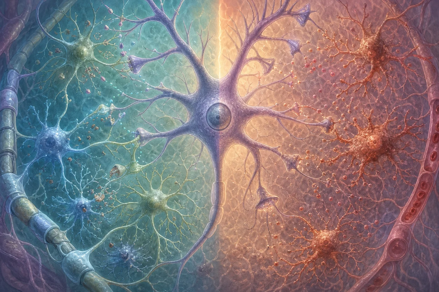
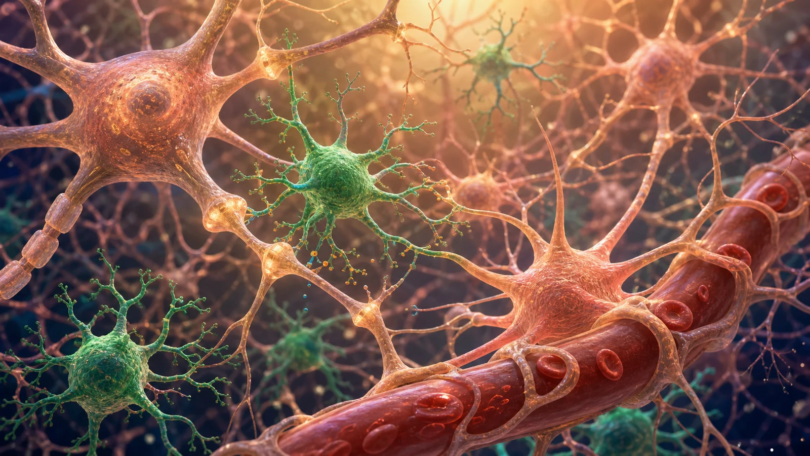
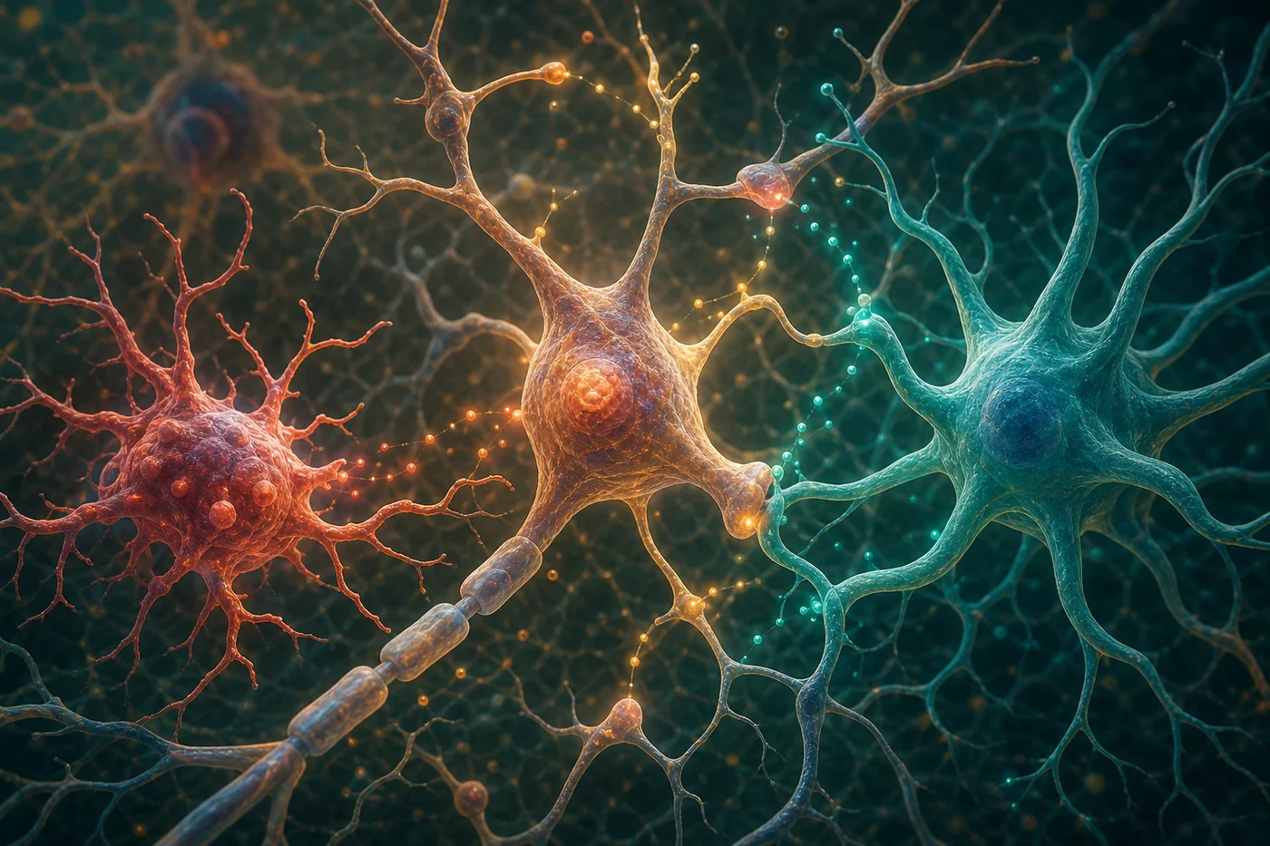
When people hear “inflammation,” they often imagine something simple: swelling, heat, danger, and a need to block it. In the brain, the story is much more interesting. Microglia and astrocytes are not temporary visitors. They are resident cells that help maintain tissue, support synapses, respond to injury, clear debris, regulate metabolism, and communicate with neurons.
In Alzheimer’s disease, those supportive roles can shift. The question is not whether inflammation is good or bad. The better question is what kind of cellular response is happening, where it is happening, when it is happening, and whether it is helping the tissue recover or adding to stress.
Microglia: Surveyors, Cleaners, and Signal Senders
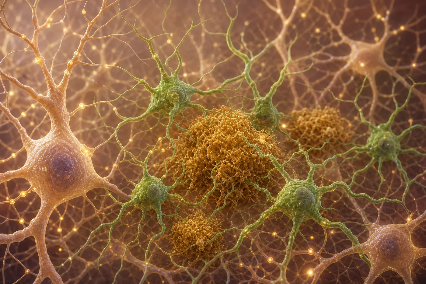
Microglia constantly survey the brain environment. Around amyloid plaques, they can gather in local neighborhoods and change the structure of nearby tissue. A Nature Communications study described how microglia can form a barrier around plaques in model systems, suggesting that some immune responses may limit damage rather than simply create it.
But microglia can also produce inflammatory signals, prune synapses, and interact with tau-related stress. Their behavior depends on context. Genetics, age, vascular health, amyloid, tau, and metabolic stress may all influence how microglia respond. That is why our articles on tau and microglia and iPSC cell models belong beside this one.
Astrocytes: Support Cells With Real Power
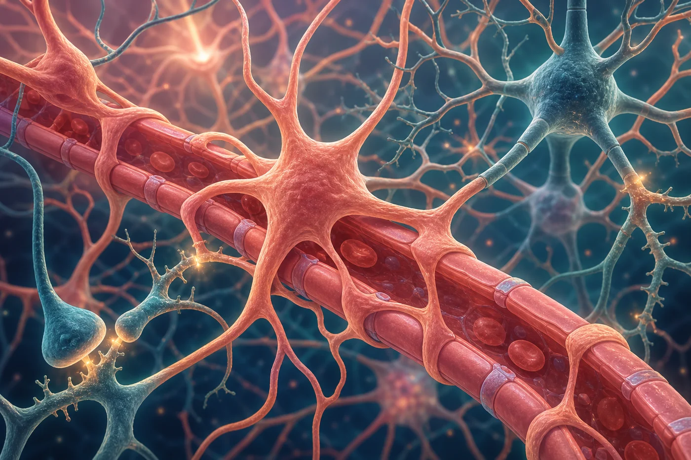
Astrocytes help regulate neurotransmitters, maintain the chemical environment around synapses, support energy metabolism, and interact with blood vessels at the blood-brain barrier. They are not background filler. They are active partners in the brain’s everyday stability.
In a widely discussed Nature paper, researchers showed that microglial signals could induce a reactive astrocyte state in disease models. Later work, including human and model-system studies, has tried to move beyond one label and understand the many states astrocytes can occupy. A 2024 Nature Neuroscience study mapped astrocyte transcriptomic changes along the progression of Alzheimer’s disease, adding detail to how astrocytes can occupy different states across space and time.
Cell States Are Not Personality Types
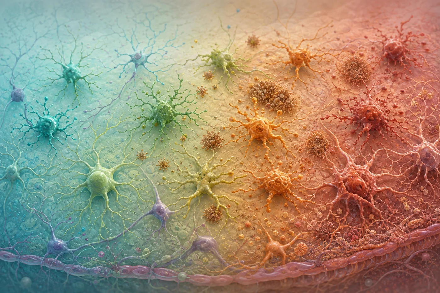
It can be tempting to label cells as protective or harmful, but biology resists that shortcut. A microglial response that clears debris early may become damaging if it stays activated too long. An astrocyte response that walls off injury may become a problem if it disrupts synaptic support. Timing changes meaning.
Single-cell and spatial methods are helping researchers see that glial states are diverse. Human work on microglial states in Alzheimer’s disease shows why the field is moving away from broad categories and toward more precise maps of cell behavior. That precision matters because future therapies may need to tune a response rather than silence it.
Why This Matters for People, Not Just Laboratories
For readers, the practical lesson is humility and hope without hype. Alzheimer’s disease is not just amyloid, not just tau, not just inflammation, and not just aging. It is a changing tissue environment. Brain immune cells may help explain why one person’s disease progresses differently from another’s and why prevention research often points back toward vascular, metabolic, sleep, and inflammatory health.
This connects with Mindful Diabetes because metabolic health is part of the body’s inflammatory and vascular landscape. That does not mean diabetes causes every Alzheimer’s case. It means the systems we care for in diabetes education also matter for brain resilience. Our guide, sleep article, blood biomarker article, and health tools are different doors into the same larger goal: helping people understand their bodies with clarity instead of fear.