
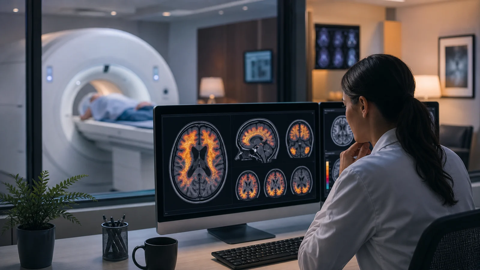
Why Seeing Tau Matters
Alzheimer’s disease is often described through plaques and tangles, but families live with symptoms: memory changes, language changes, navigation problems, confusion, and loss of independence. Tau PET imaging matters because tau patterns often line up more closely with symptoms and neurodegeneration than amyloid alone. It gives researchers a way to see where tau-related biology is appearing in the living brain.
That does not make tau PET a perfect crystal ball. It is an imaging tool with technical limits, cost barriers, tracer differences, and interpretation rules. But by October 13, 2025, tau imaging had become one of the most important ways researchers connect molecular pathology to the clinical picture.
Amyloid PET and Tau PET Ask Different Questions
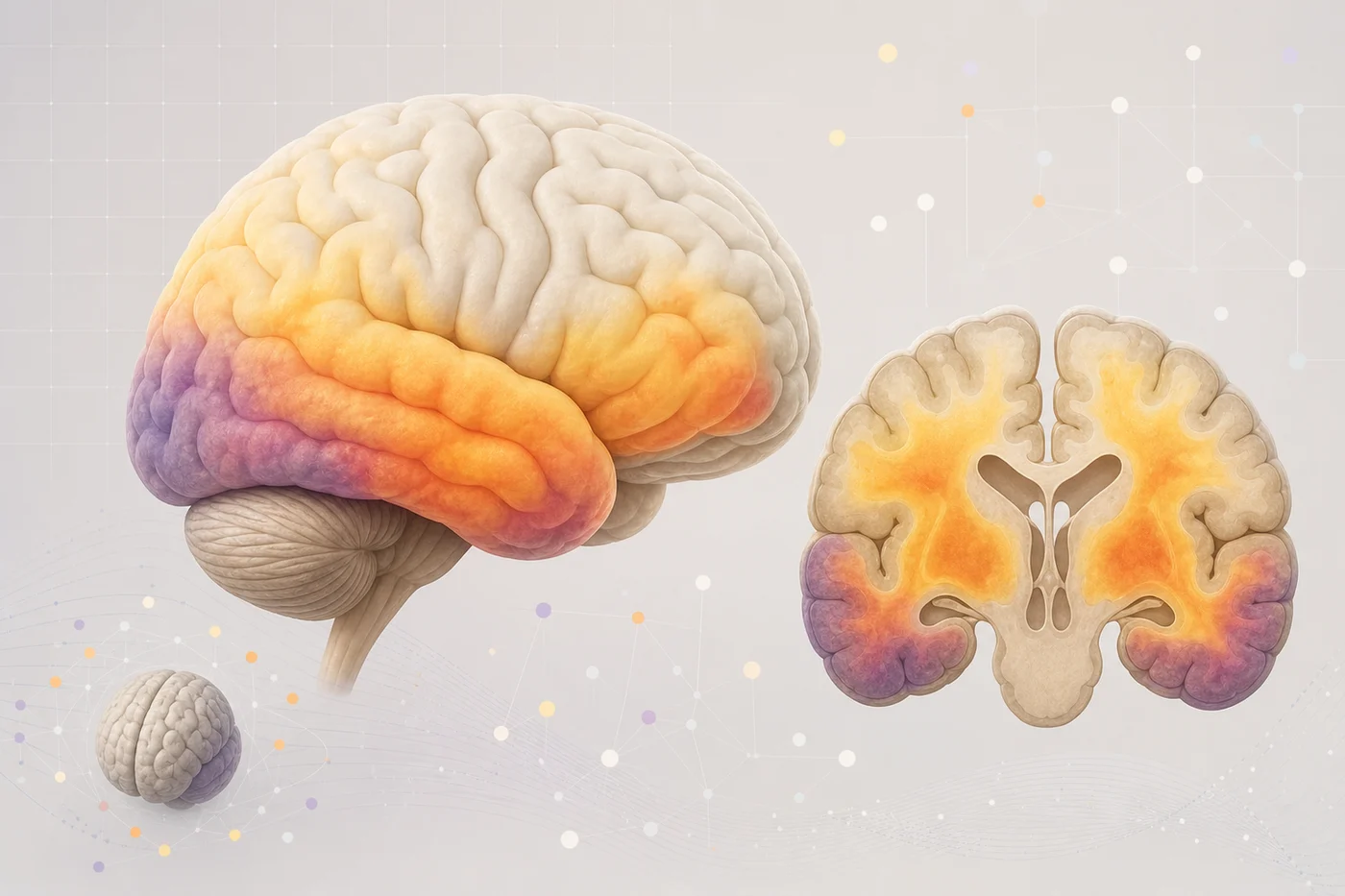
Amyloid PET can help show whether amyloid plaque pathology is present. Tau PET asks more about where tau has accumulated and how that pattern may relate to disease stage. The updated appropriate use criteria for amyloid and tau PET reflect this more careful approach: imaging should answer a clinical or research question, not be ordered just because a technology exists.
For readers following our Alzheimer’s series, this connects directly to amyloid research and tau biology. Amyloid may set the stage in many cases. Tau may track the brain networks that are becoming stressed. Both are part of the story, and neither is the whole story.
Regional Patterns and Symptoms
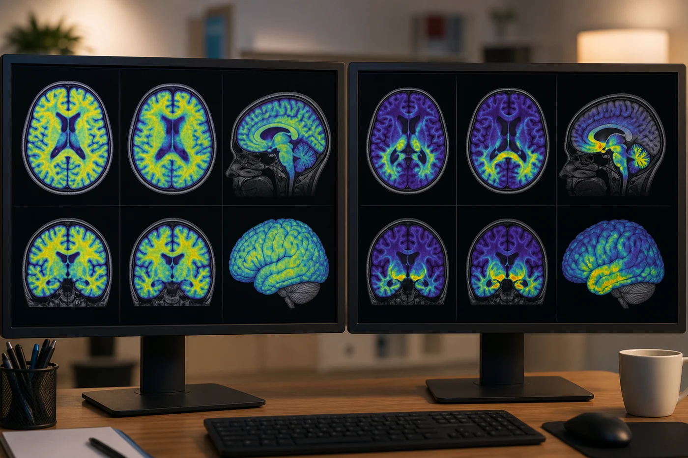
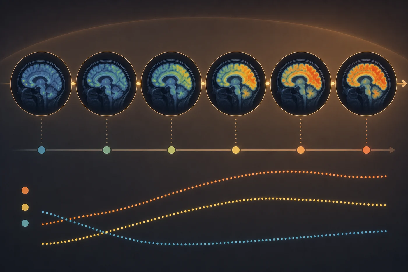
One of the powerful features of tau PET is its regional detail. A scan can show whether signal appears in medial temporal areas, association cortices, or other patterns that researchers can compare with cognitive testing and structural brain change. Earlier research linked tau PET patterns with clinical impairment and cognition, including work summarized in human tau imaging studies.
That regional view helps explain why two people with the same broad diagnosis can look different day to day. Alzheimer’s is not only a chemistry problem. It is a network problem. The pattern of affected circuits can influence which symptoms appear first and which supports a family may need.
The Limits of the Window
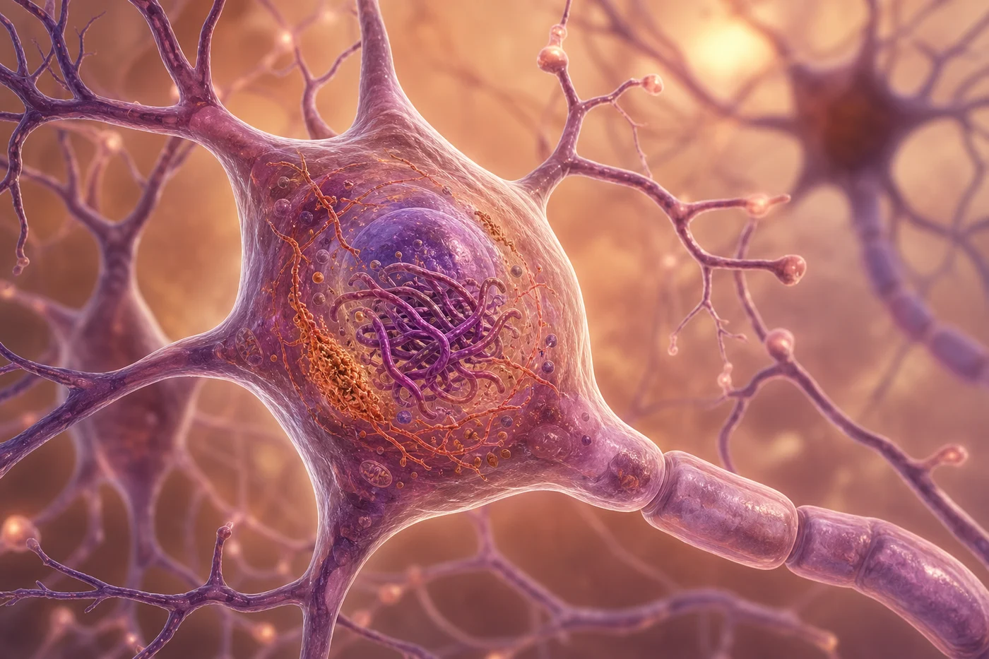
Tau PET is still a window, not the room itself. Tracers can differ. Some can bind off-target in regions that complicate interpretation. A tau PET tracer review discusses why tracer choice and technical details matter. Access is also uneven. PET imaging can be expensive, and not every community has a center that can perform or interpret it.
The Tauvid label is a reminder that imaging tools come with specific indications and limitations. A scan should support a thoughtful clinical process. It should not turn uncertainty into a false sense of certainty.
Where Blood Biomarkers Fit
Blood biomarkers may eventually help decide who needs tau PET, who should be referred to a specialist, or who may qualify for a particular study. They may also reduce inequity if they are validated and used responsibly. Our article on Alzheimer’s blood biomarkers explores that promise and caution.
The future may be multimodal: symptoms, family history, cognitive testing, MRI, blood markers, amyloid tools, tau imaging, and longitudinal follow-up. That sounds complicated because the brain is complicated. The gift of better tools is not that they make Alzheimer’s simple. It is that they may help us be more specific, more honest, and more useful.